About NeuroSkeletal Imaging
In 2004, Neuroskeletal Imaging, fondly known as NSI, was founded and incorporated on an ideal that centered on patient care and satisfaction. We believe that outpatient radiological facilities should be owned and operated by board-certified and licensed radiologists, which allows for non-biased medical care to be efficiently coordinated with physicians and other medical providers.
Services

3.0 Tesla MRI
Magnetic Resonance Imaging (MRI) is a very safe, comfortable, advanced imaging technology.

1.5 Tesla Open MRI
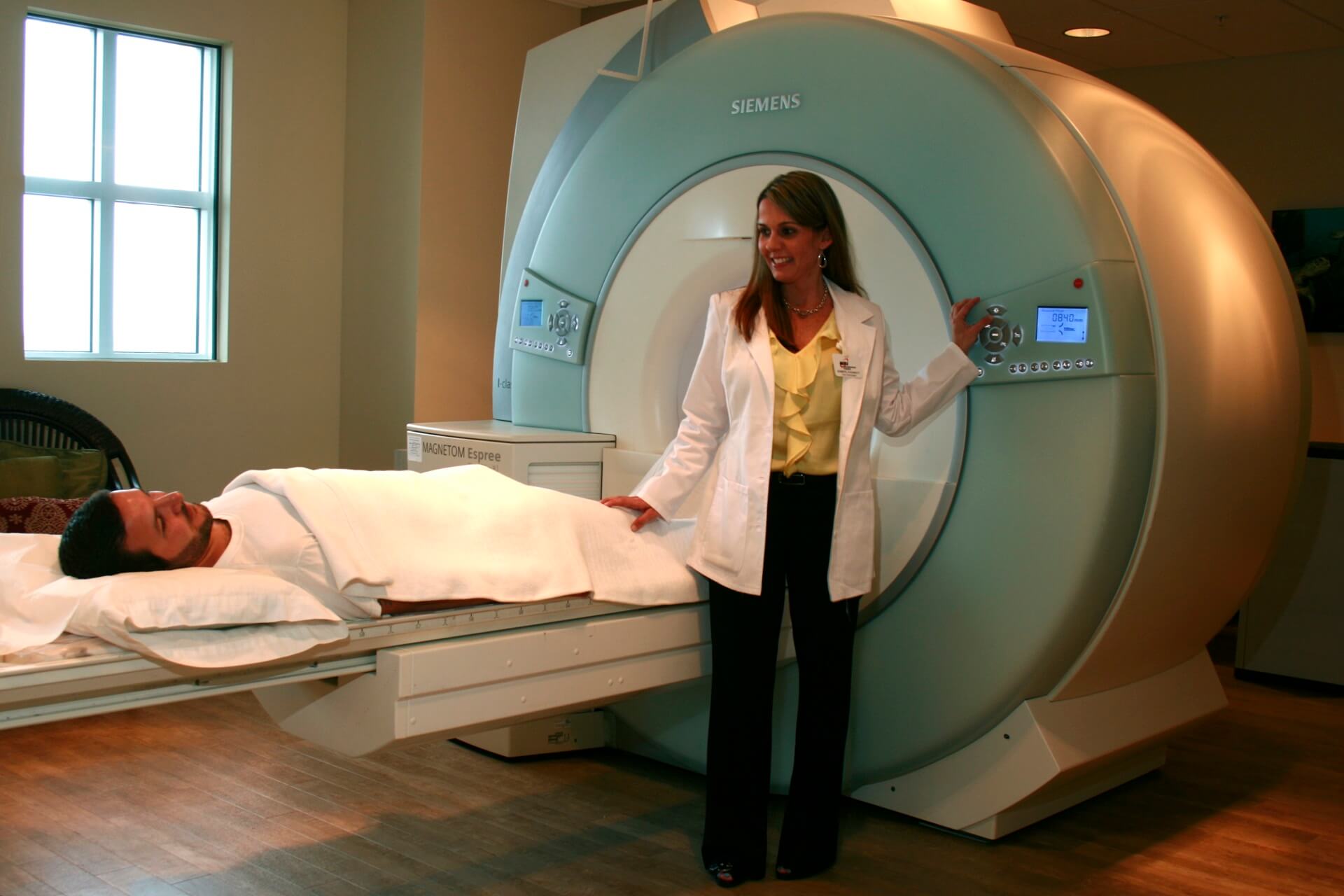
Our Siemens MAGNETOM Espree High-Field 1.5T Open MRI is the only open bore (tube) MRI

Traumatic Brain Injury Protocol

MRA & CTA
Magnetic Resonance Angiography (MRA) is an MRI technique used to image blood vessels.

16 & 64 Slice CT
Computed Tomography, known as CT, is a diagnostic procedure that uses special x-ray equipment

Arthrogram
An arthrogram is a procedure in which a radiologist, using x-ray guidance (fluoroscopy)

Ultrasound
Ultrasound imaging, also referred to as sonography, is a painless
procedure

Digital X-Ray
X-ray is one of the oldest forms of radiology testing. It is a fast and painless test..

Sedation
Sedation is a means of lowering
the anxiety level and somewhat
depressing the state

DEXA
(DEXA) Dual-energy x-ray absorptiometry is a technique used to aid in the diagnosis of osteopenia and osteoporosis. DEXA is currently available in our Merritt Island location.

Low Dose CT
The low-dose spiral CT scan continuously rotates in a spiral motion and takes several 3-dimensional X-rays of the lungs. These X-rays are very detailed and can show early-stage lung cancers..
Locations
MERRITT ISLAND
255 North Sykes Creek Parkway, Suite 102, Merritt Island, Florida 32953
(321) 454-4897

Reviews & Awards

Frequently Asked Questions
What is an MRI?
How does MRI work?
A large magnet surrounding the body causes the hydrogen nuclei (protons) in water molecules to align in a particular direction, and then radio waves strike the protons, causing them to spin out of alignment. When the radio signal is turned off, the protons return to their previous alignment, giving off new radio waves at the same time. The returning radio signals are received and analyzed by a computer, which then creates cross-sectional images of the body.
When is MRI used?
- Brain and nervous system: MRI is the most accurate tool for evaluating most diseases of the brain and spinal cord, including strokes, tumors, and multiple sclerosis.
- Musculoskeletal system: MRI is the most accurate way to non-invasively image most joints (knees, shoulders, wrists, etc) for problems involving cartilage, ligaments, tendons and muscles.
- Breast: with our new EXCITE technology upgrade come H.D. VIBRANT (Volume Imaging for Breast Assessment). We can now simultaneously examine both breasts in high resolution in a single patient visit.
- Other: MRI can also be useful for evaluating the neck, liver, kidneys, pelvic organs, blood vessels, and lymph nodes.
What is MRA?
What is a CT or CAT scan?
When is CT used?
- Head & Neck: CT can be used to evaluate the brain for strokes, headaches, or injury. It can also be used to evaluate the sinuses, facial bones and structures of the neck.
- Chest: CT is excellent for evaluating the lungs for infection, emphysema, tumors and other conditions.
- Abdomen & Pelvis: CT is extremely useful for visualizing all organs of the abdomen and pelvis, and is often performed to evaluate the possibility of appendicitis, diverticulitis, kidney disease, liver or gallbladder disease, etc.
- Spine: CT is very helpful in evaluation of spinal fractures, and can also be used to evaluate disc herniations and other causes of neck and back pain.
- Blood vessels: A type of CT scan can be performed for visualization of arteries or veins; it is called CT Angiography (CTA). The latest multidetector CT scanners produce the most accurate CTA examinations.

